Published on Oct. 19, 2020
Avian Metapneumovirus in poultry
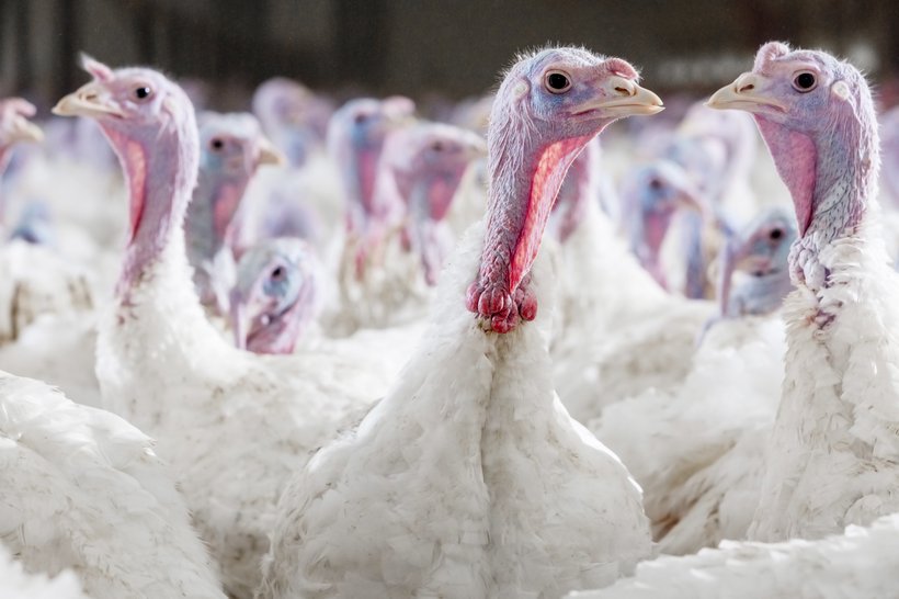
Avian Metapneumovirus (aMPV) infections affect several species of domestic poultry and causes upper respiratory tract disease as TRT (turkey rhinotracheitis) in turkeys and SHS (swollen head syndrome) in chickens, as well as reproductive disorders. In layers aMPV has also economic importance since infection leads to significant drops in egg production and deterioration of egg quality. Severity of the disease will depend on the presence of bacterial secondary infections and environmental distress. Brown egg layers are more susceptible to the disease than white egg layers.
aMPV are currently divided into four subtypes (A, B, C and D), genetically different and with variable prevalence in different areas of the world. Although the disease is highly contagious, the virus does not survive for long in the environment and it is easily inactivated by most common disinfectants.
Transmission is by direct contact and the airborne route. The virus replicates mainly in epithelial cells of the upper respiratory tract (sinuses, trachea, nasal turbinates), damaging the cilia and allowing easier secondary pathogens infection. Virus can also be isolated from reproductive tract but there is no evidence of vertical transmission.
Clinical Signs
In layers, infection is not always associated with clinical signs: unspecific respiratory symptoms can appear, nasal distillation, watery eyes, sneezes and rarely “swollen head”, depending if other predisposing factors like poor ventilation or other pathogens (Mycoplasma, E. coli, virus with respiratory tropism) are present. Drop in egg production, pale brown eggs and poor eggshell quality are frequently the only signs observed.
Layer flocks seem to be more susceptible to aMPV by the peak of production, but cases have been reported at any age.
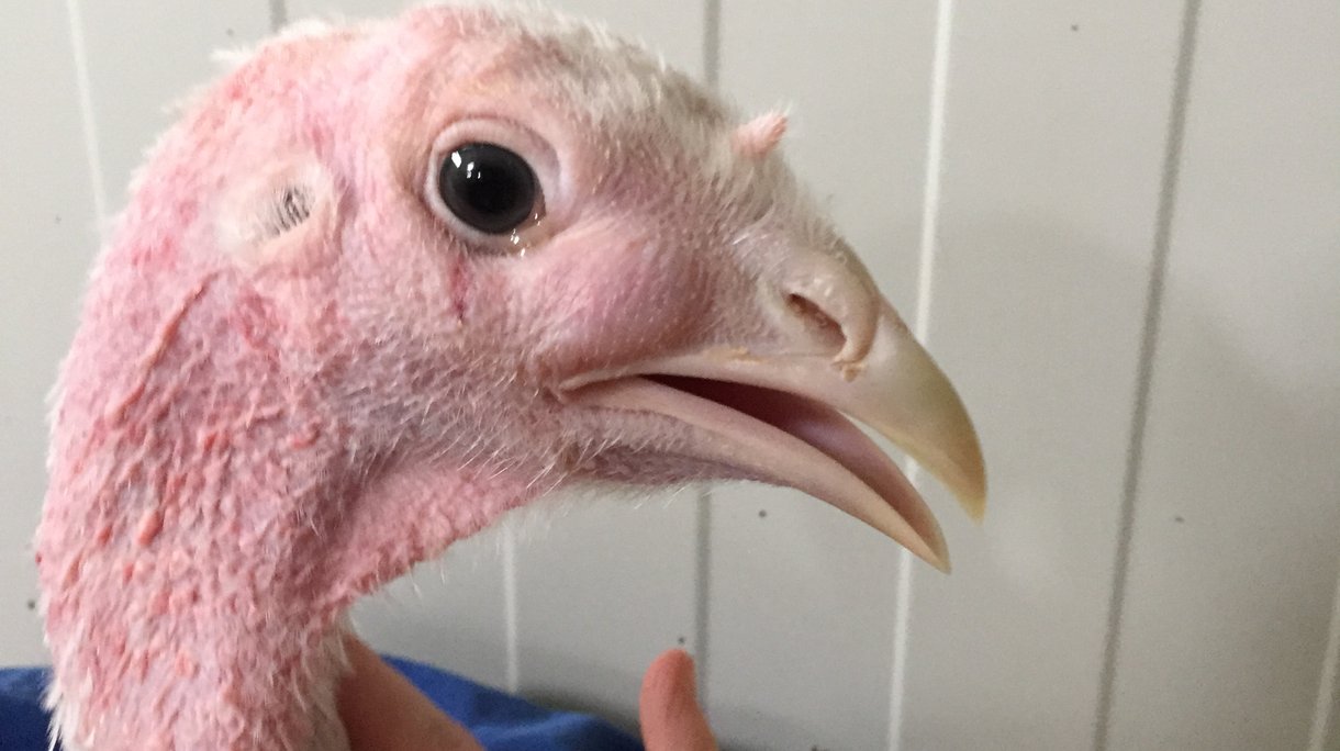
Severity of gross lesions depend mostly on secondary infection: from light congestion to severe oedema with presence of mucus in nasal cavity in sinuses, ovary regression and even peritonitis. The most relevant microscopic lesion is loss of cilia or paralyse of cilia activity of epithelial cells from the upper respiratory tract.
Diagnosis
Clinical signs are not specific, therefore diagnosis should be made by virus isolation, RT-PCR or ELISA. Samples of choice for virus detection are tissues from the upper respiratory tract or swabs from the same origin, ideally from early stages of the disease and even from apparently healthy birds from the suspected flock.
ELISA tests can also be used but sensitivity can vary depending on subtype antigens used.
Control & Vaccination
There is no effective treatment for aMPV infection, but if necessary, treatment with antibiotics could help to control secondary bacteria.
Prevention strategy should be based not only in vaccination but also in strict biosecurity and proper flock management practices to minimize predisposing factors: optimal ventilation (avoiding dust and ammonia), suitable stocking density, proper temperature control etc.
Live attenuated vaccines administered by spray or drinking water are available for immunization of young pullets in rearing and are essential for stimulating local immunity and blocking the entrance of the virus. Combination with inactivated vaccines after priming with live vaccines is recommended for stronger and longer-lasting protection.
Cell-mediated immunity plays a more decisive role in protection against aMPV than humoral immunity and antibody titres induced by vaccination are not considered a very sensitive indicator of protection.
References
- Picture affected turkey: Dr. Rebecca Lindenwald